Gelatin and casein zymography
Zymography is a specialised form of protein electrophoresis used to detect and compare matrix metalloproteinase (MMP) enzymes in samples such as conditioned medium, tissue extracts, or serum. Enzymes are separated by size in a substrate-embedded polyacrylamide gel, renatured after electrophoresis, and visualised as clear bands where the embedded gelatin or casein has been digested.
What is zymography?
Enzymes in the sample are separated by size exactly as in normal SDS-PAGE. Once electrophoresis is complete, the proteins are renatured so they can regain proteolytic activity. They then digest a substrate protein embedded in the gel during polymerisation. Upon staining with a protein stain such as Coomassie Blue, protease bands appear as clear regions of substrate lysis against a blue background.
Unlike a standard SDS-PAGE gel, which shows proteins stained with a general protein stain, zymography uses the enzyme’s own activity to reveal whether those proteins are present in a sample.
A very important point to understand is that although the technique uses inherent enzyme activity for detection, zymography does not measure MMP activity in the original sample. Many MMP enzymes are secreted as inactive pro-MMPs. Because these become partially denatured by SDS during electrophoresis, they auto-activate during the renaturation step. They are detected by activity, but they were not necessarily active in the original sample. Even if an MMP is in an active form (pro-peptide cleaved), it may not be active in the sample because of tissue inhibitors of metalloproteinases (TIMPs). MMP–TIMP complexes separate during electrophoresis, so the enzyme becomes active only at the renaturation step.
Gelatin vs casein zymography
The two most common forms of zymography are:
- Gelatin zymography — used mainly to study gelatin-degrading enzymes such as MMPs, especially MMP-2 (gelatinase-A) and MMP-9 (gelatinase-B).
- Casein zymography — used to detect enzymes that degrade casein. It is commonly used for certain MMPs, mainly MMP-3 (stromelysin) but also MMP-2 and MMP-9, as well as serine proteases and other casein-degrading enzymes.
The basic principle
A protein substrate (gelatin or casein) is mixed directly into the polyacrylamide gel mix before it polymerises. Samples such as cell culture conditioned medium, mixed with SDS sample buffer, are loaded and separated by electrophoresis. It is critical that this buffer does not contain a reducing agent such as β-mercaptoethanol or DTT, and the sample must not be heated before loading.
After electrophoresis, SDS must be removed from the gel so enzymes can refold. This is done by incubating the gel in buffer containing Triton X-100. Triton X-100 forms micelles with a hydrophobic core that attracts SDS hydrophobic tails. Once SDS is removed, the gel is incubated in a buffer that allows MMPs to digest the embedded substrate. Finally, the gel is stained to reveal areas where substrate has been digested, which appear as clear bands against a dark blue background.
These clear bands indicate the presence of proteases. Remember that both active and pro-MMPs can be detected.
Method
Step 1: Prepare a gel containing substrate
Cast an SDS-PAGE gel with gelatin or casein incorporated into the matrix. Gelatin acts as the substrate that proteases digest. Gelatin can be purchased from suppliers such as Merck; we often obtained better results using freshly denatured collagen boiled in water. We use gelatin at 2 mg/ml in a 7.5% polyacrylamide gel mix. For casein zymography we use 4 mg/ml β-casein.
Step 2: Load samples
Samples may include cell culture supernatants, tissue extracts, or serum/plasma. Unlike standard SDS-PAGE, samples are not boiled and reducing agents are omitted because they can permanently inactivate proteases. Use a non-denaturing loading buffer such as 50 mM Tris pH 6.8, 10% glycerol, 4% SDS, and 0.025% bromophenol blue.
Step 3: Electrophoresis
Proteins are separated according to molecular weight. Although SDS partially denatures the enzymes, they remain capable of being reactivated later.
Step 4: Renaturation
Soak the gel in buffer containing 2.5% Triton X-100 (for example, 2 × 15 min) to remove SDS and allow proteases to refold.
Step 5: Enzyme development
Incubate the gel in activation buffer (often containing calcium and zinc ions for MMPs), for example 50 mM Tris pH 8.0, 50 mM NaCl, 10 mM CaCl2, 0.05% Brij 35. During this incubation, active proteases digest the gelatin or casein in their immediate vicinity. The amount of digestion depends on enzyme activity and incubation time.
Step 6: Staining
Stain with Coomassie Blue. Intact gelatin or casein stains dark blue; degraded regions do not retain stain and appear as clear bands.
To confirm that bands of lysis are due to MMPs rather than other proteinases, run a positive sample across multiple lanes, cut the gel into strips after SDS removal, and incubate each strip in development buffer with different protease inhibitors (for example AEBSF, NEM, BB-94, or EDTA as a divalent metal ion chelator). If EDTA and an MMP inhibitor block band formation but AEBSF or NEM do not, the bands are likely due to MMP action.
Interpretation
- Clear band = active substrate-degrading enzyme.
- Band position = approximate molecular weight (pro-MMP-9 @ 95 kDa, active MMP-9 @ ~85 kDa, pro-MMP-2 @ 72 kDa, active MMP-2 @ 68 kDa, stromelysin @ 50 kDa).
- Band intensity = relative enzyme abundance (semi-quantitative).
How casein zymography works
Casein zymography follows essentially the same workflow, except the gel contains casein instead of gelatin. Some proteases, such as stromelysin, digest casein more efficiently than gelatin, so casein zymography can reveal enzymes that are difficult to detect with gelatin alone.
What information does zymography provide?
- Whether a protease is present in pro- or processed form.
- The approximate molecular weight of the protease.
- Relative differences in enzyme abundance between samples.
Advantages
- Extremely sensitive.
- Detects enzyme activity directly.
- Relatively inexpensive.
- Can distinguish different proteases by molecular weight.
Limitations
- Mostly semi-quantitative rather than fully quantitative.
- Only detects enzymes that can renature after electrophoresis.
- Different enzymes may migrate similarly, making identification difficult.
- Additional methods (western blotting, inhibitor studies, mass spectrometry) are often needed to confirm enzyme identity.
Quick comparison
| Feature | Gelatin zymography | Casein zymography |
|---|---|---|
| Substrate in gel | Gelatin | Casein |
| Common targets | MMP-2, MMP-9 | Various MMPs and serine proteases |
| Readout | Clear bands in gelatin background | Clear bands in casein background |
| Main purpose | Detect gelatin-degrading MMPs | Detect casein-degrading MMPs (e.g. MMP-3) |
Examples
This is a gelatin zymogram showing MMP-9 bands at 95 kDa and MMP-2 bands at 72 kDa. It was made by running cell culture media samples from cells cultured overnight with combinations of growth factors and cytokines.

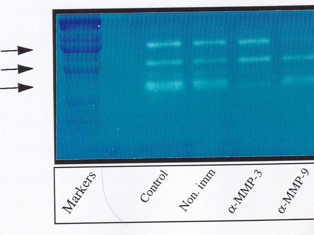
This is an example of casein zymography showing bands at 95 kDa (MMP-9), 72 kDa (MMP-2), and 50 kDa (MMP-3). Before electrophoresis, MMP-9 or MMP-3 were immunoprecipitated from the indicated samples to confirm band identity.

This is another example of casein zymography where the same sample was run in all lanes. The gel was cut into strips after electrophoresis and incubated overnight with the indicated protease inhibitors. Ro-31-9790 is an MMP-2 inhibitor. Note the absence of bands in strips incubated with Ro-31-9790 and EDTA, indicating the bands are the result of MMP activity.
